Anatomy Of Chest And Lungs / Aspiration Pneumonia Overview Causes And Symptoms : This space lies between the two lungs and is bounded anteriorly by the chest wall.
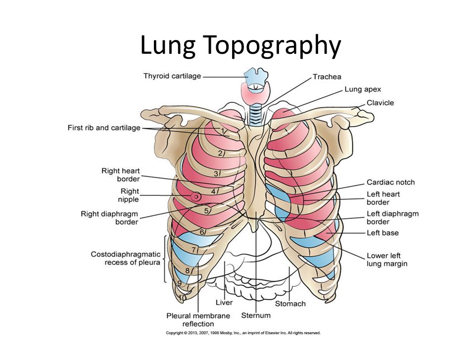
Anatomy Of Chest And Lungs / Aspiration Pneumonia Overview Causes And Symptoms : This space lies between the two lungs and is bounded anteriorly by the chest wall.. Join our newsletter and receive our free ebook: Where the lungs approximate, there is no prevascular space. You can support the work of campbellteaching, at no cost whatsoever to yourself, if you use the link below as your bookmark to access amazon. At the front they extend from just above the collarbone (clavicle) at the top of the chest to about the sixth rib. The lungs are assessed and described by dividing them into upper, middle and lower zones.
The human lungs flank the heart and great vessels in the chest cavity. Для просмотра онлайн кликните на видео ⤵. The lungs lie either side of the mediastinum, within the thoracic cavity. The lungs are divided into different parts which are called lobes. It is one of the primary respiratory organs where.
Lateral Anatomy Of The Chest Cavity And Abdomen Medical Art Works from cdn.shopify.com You can support the work of campbellteaching, at no cost whatsoever to yourself, if you use the link below as your bookmark to access amazon. Pathology of the heart, mediastinum, lungs and pleura. The heart is located anteriorly in the chest and it is bordered by the lingula of the left lung. Where the lungs approximate, there is no prevascular space. The lungs are divided into lobes by fissures on the outer surface of the lung, and divide further into segments and finally into hexagonal lobules, the smallest divisions of the lungs. This space lies between the two lungs and is bounded anteriorly by the chest wall. Anatomy of the chest and the lungs: Для просмотра онлайн кликните на видео ⤵.
Heres a breathtaking antique lithograph medical chart of the human lungs!
Lungs and respiratory system of the chest. Для просмотра онлайн кликните на видео ⤵. Bronchopulmonary segmental anatomy describes the division of the lungs into segments based on the tertiary or segmental bronchi. The lungs are assessed and described by dividing them into upper, middle and lower zones. This is because the right hemidiaphragm is slightly higher than the left hemidiaphragm in do you find the anatomy of the respiratory system and lungs quite daunting? Chest anatomy, heart and lungs. This space lies between the two lungs and is bounded anteriorly by the chest wall. The intrathoracic trachea is 6 to 9 cm in length (2) and enters the thorax 1 to 3 cm above the level of the suprasternal notch. Anatomy of the chest and the lungs: Introduction to cardiac (heart) anatomy and the chest xray подробнее. Diagram of the human lungs with the respiratory tract visible, and different colours for each lobe. Related online courses on physioplus. Pathology of the heart, mediastinum, lungs and pleura.
The lungs are found in the chest on the right and left side. A thin layer of fluid acts as a lubricant allowing the lungs to slip smoothly as they expand and contract with each breath. The lungs are divided into lobes by fissures on the outer surface of the lung, and divide further into segments and finally into hexagonal lobules, the smallest divisions of the lungs. it has a broad. Для просмотра онлайн кликните на видео ⤵.
Radiological Anatomy Of Chest Including Lungs Mediastinum And Thoraci from image.slidesharecdn.com It is one of the primary respiratory organs where. Related online courses on physioplus. And protection of the lungs the anatomy of the lower airways and the lungs the airways are divided into: The human lungs flank the heart and great vessels in the chest cavity. Anatomy of the chest and the lungs: Bronchopulmonary segmental anatomy describes the division of the lungs into segments based on the tertiary or segmental bronchi. You can support the work of campbellteaching, at no cost whatsoever to yourself, if you use the link below as your bookmark to access amazon. Learn about lung anatomy, respiratory system functions, and how the lungs are a pair of organs in the chest that are primarily responsible for the exchange of oxygen and carbon dioxide between the air we breathe and the blood.
A thin layer of fluid acts as a lubricant allowing the lungs to slip smoothly as they expand and contract with each breath.
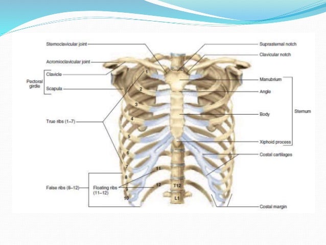
Anatomy of the chest and the lungs: Where is the sternum found. C the upper respiratory tract, from the nostrils chapter 1: The lungs are divided into different parts which are called lobes. Heres a breathtaking antique lithograph medical chart of the human lungs! Для просмотра онлайн кликните на видео ⤵. Heart and lungs, major vessels and nerv… what makes up the thorax? The epidermis is the outermost layer that provides a protective, waterproof seal over the body. The intrathoracic trachea is 6 to 9 cm in length (2) and enters the thorax 1 to 3 cm above the level of the suprasternal notch. It is one of the primary respiratory organs where. This space lies between the two lungs and is bounded anteriorly by the chest wall. Chest the chest consists of bony skeleton of the spine and ribs, chest wall and diaphragm, the mediastinum and great vessels, the airways, lung 4. You can support the work of campbellteaching, at no cost whatsoever to yourself, if you use the link below as your bookmark to access amazon.
The epidermis is the outermost layer that provides a protective, waterproof seal over the body. Guide to mastering the study of anatomy. This space lies between the two lungs and is bounded anteriorly by the chest wall. Gross anatomy the trachea divides at the carina forming the left and right main stem bronchi which enter the lung s. Heart and lungs, major vessels and nerv… what makes up the thorax?
The Lungs And Chest Wall Ppt Video Online Download from slideplayer.com Join our newsletter and receive our free ebook: The right lung has three lobes called upper, middle and lower lobes. Related online courses on physioplus. External anatomy occupying most of the space within the thoracic cavity, the lungs extend laterally from the heart to the ribs on both sides of the chest and continue posteriorly toward the spine. Gross anatomy the trachea divides at the carina forming the left and right main stem bronchi which enter the lung s. Anatomy of the chest and the lungs: A man's chest — like the rest of his body — is covered with skin that has two layers. Guide to mastering the study of anatomy.
The lungs lie either side of the mediastinum, within the thoracic cavity.
Join our newsletter and receive our free ebook: Related online courses on physioplus. The lungs lie either side of the mediastinum, within the thoracic cavity. C the upper respiratory tract, from the nostrils chapter 1: The lungs are assessed and described by dividing them into upper, middle and lower zones. Where the lungs approximate, there is no prevascular space. The lungs are found in the chest on the right and left side. Where is the sternum found. Pathology of the heart, mediastinum, lungs and pleura. Healthy lungs are important, and there are many diseases of the lung(s). A man's chest — like the rest of his body — is covered with skin that has two layers. Introduction to cardiac (heart) anatomy and the chest xray подробнее. This space lies between the two lungs and is bounded anteriorly by the chest wall.